Advanced Dental Technology – Jersey City, NJ
Ensuring Greater Accuracy with the Help of Innovative Technology

At North Jersey Endodontic Group of Jersey City, our specialists incorporate the use of modern dental technology to ensure greater accuracy and precision when treating patients with tooth pain. Using an array of equipment that includes digital dental X-rays, a CT/cone beam scanner, a laser-activated irrigation system, and a dental operating microscope, we can pinpoint even the smallest problem areas before crafting personalized treatment plans that are designed to eliminate the issue and restore oral health.
CT/Cone Beam Scanner
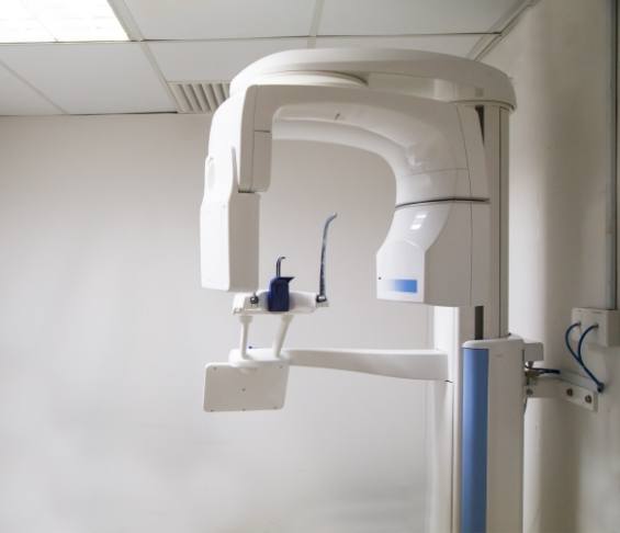
Our CT/cone beam scanner is a unique device that uses 3D technology to allow for enhanced imaging and accurate treatment planning. Rotating a full 360 degrees around your head, it captures pictures of your oral and facial structures, generating a three-dimensional model that our team can use to prepare for root canal treatment. Not only can we see your teeth, gums, jawbone, blood vessels, and nerve pathways, but we can indicate a precise plan that ensures positive results.
Dental Operating Microscope
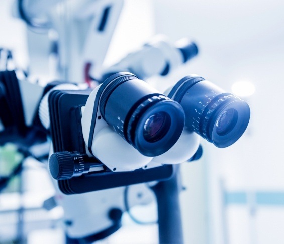
When it comes to identifying the problem area, it can often be hard to see certain parts of a tooth. Even the most enhanced imaging systems may not be enough. However, with our dental operating microscope, our endodontist’s vision is enhanced by up to 25 times, making it possible to detect even the smallest areas of bacteria that need to be removed when performing a root canal.
Digital Radiography

Traditional radiographs were once the only way for dentists and specialists to see what was happening beneath the gumline. However, they often required uncomfortable methods and longer appointments because of how long it took for the images to develop. Fortunately, our team uses digital X-rays that cut down on treatment time and produce higher-resolution images that allow us to better analyze your oral structure when preparing your treatment plan.
About Us Meet the Dentists Meet the Team Educational Videos View Our Services